Most diseases causing elevation of the liver enzymes don’t necessarily mean alternation of the liver in abdominal ultrasound.
Possible causes of elevated liver enzymes and negative ultrasound include:
- Some cases of nonalcoholic fatty liver disease (NAFLD).
- Alcoholic hepatitis.
- Viral hepatitis (particularly hepatitis B and C).
- Toxins and drugs that cause elevated liver enzymes.
- Autoimmune hepatitis.
- Others such as primary biliary cholangitis, Wilson’s disease, hemochromatosis, etc.
1. Non-alcoholic fatty liver disease (NAFLD).
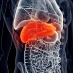
Non-alcoholic fatty liver disease is an abnormal accumulation of excess fat inside the liver cells. It is one of the most common diseases worldwide.
NAFLD (non-alcoholic fatty liver disease) is subdivided into:
- NAFL (non-alcoholic fatty liver): the accumulation of fat inside the liver cells without inflammation (elevation of the liver enzymes).
- NASH (non-alcoholic steatohepatitis): Occurs when fat accumulation inside the liver leads to inflammation (elevation of the liver enzymes).
At least one in every four people (25%) has fatty liver (NAFLD). And that’s why it came on the top list of the causes of elevated liver enzymes with negative ultrasound.
The table below illustrates the prevalence of fatty liver (NAFLD) in different regions.

Why does negative ultrasound not exclude fatty liver?
Typically, The fatty liver appears slightly brighter than normal on ultrasound. However, it is not the case for all patients with NAFD.
- The overall sensitivity of ultrasound in detecting NALFD is 85%. So, at least 15% of people with NAFLD or NASH will have a negative ultrasound (reference).
- The liver ultrasound examination is even less accurate in patients with significant obesity. The sensitivity of ultrasound may be less than 50% (reference).
What causes NAFLD?
A combination of factors causes NAFLD. Most commonly, a combination of dietary and genetic factors.
Here are the most common risk factors for fatty liver (reference):
- Obesity.
- Diabetes (type two).
- Hypertension.
- Dyslipidemia (increased lipids such as cholesterol and triglycerides).
- Males are more likely to develop NASH than females.
- Age also plays a role, the prevalence of NAFLD increases with age.
- Race Hispanics have the highest fatty liver (NAFLD) prevalence, followed by Caucasians and African Americans.
How is NAFLD diagnosed?
To diagnose NAFLD, your doctor has to fulfill all the following:
- Confirmation of liver steatosis (fat accumulation) by imaging or biopsy.
- Exclusion of significant alcohol consumption.
- Exclusion of other causes of fat accumulation in the liver (such as lipodystrophy, hepatitis C, starvation, acute fatty liver of pregnancy, etc.).
- Absence of coexisting liver diseases.
To confirm fat accumulation in the liver, your doctor may visualize it by abdominal ultrasound, CT, MRI, or transient elastography.
As we explained before, NAFLD may be negative in at least 15% of people with NAFLD who will have a negative ultrasound.
The gold standard for detecting hepatic fat accumulation is taking a biopsy from your liver with a specialized needle.
But your doctor will not take a biopsy unless he excludes other causes of elevated liver enzymes (such as alcohol and medications).
Some clinicians are against the idea of liver biopsy because it is an invasive maneuver, and they prefer watchful waiting for the liver enzymes to improve.
Discuss the issue with your doctor for the best decision that suits your condition.
2. Alcoholic hepatitis.
Patients with chronic alcohol use may develop liver inflammation which manifests as elevated liver enzymes (even if the ultrasound is negative).
Consumption of significant amounts of alcohol leads to rapid fat accumulation and inflammation of the liver in a mechanism similar to NAFLD.
Alcoholic hepatitis results in both acute attacks and chronic forms. Patients with heavy alcohol use (>100 g pure alcohol per day) will develop alcoholic hepatitis.
The acute attacks may present with liver pain, jaundice, anorexia, and elevated liver enzymes. The ultrasound may be negative in acute or early stages of alcoholic hepatitis.
What is considered significant alcohol intake?
The significant amount varies with age, gender, and other coexisting diseases.
A standard drink contains 14 grams of pure alcohol, the (significant) amounts that increase the risk of alcohol-related liver inflammation and elevation of liver enzymes are:
- For men under age 65:
– More than 14 standard drinks per week on average.
– More than four drinks on any day. - For women and adults over 65:
– More than seven standard drinks per week.
– More than three drinks per day.
The illustration below shows different alcohol concentrations in various drinks (reference):

3. Viral Hepatitis (Mainly hepatitis B & C).
Viral liver infections (acute or chronic) are among the most common causes of elevated liver enzymes ultrasound is often negative in the acute and early stages of chronic viral hepatitis.
About 296 million people are living with hepatitis B worldwide. Also, more than 170 million people have hepatitis C.
Possible modes of transmission of hepatitis B and c viruses (HBV, HCV) include:
- Dental procedures.
- Transfusion of blood or blood products (such as plasma, coagulation factors, or platelets.
- Sharing needles or syringes.
- Sexual contact with an infected person (especially HBV).
- From a mother to her baby at birth (especially HBV).
Symptoms of chronic hepatitis B and C:
- The condition may be completely asymptomatic in many cases.
- Mild symptoms such as chronic fatigue, anorexia, and joint pain.
- Yellowish discoloration of the skin and eye whites.
- Dark urine, pale stool (only in acute flare-ups or end-stage disease).
- Abdominal pain.
Diagnosis:
If you have elevated liver enzymes (even with a negative ultrasound), your doctor will often screen for viral hepatitis.
Testing for the hepatitis C virus antibodies (HCB Abs) and Hepatitis B surface antigen (HBsAg) are easy and rapid screening tests for the viruses.
If one is positive, your doctor will proceed with the investigations to determine the viral load and the need for treatment.
4. Toxin- and Drug-induced liver inflammation.
Drugs and toxins are among the widespread causes of unexplained elevations of liver enzymes.
In most cases of drug- or toxin-induced liver inflammation, you may have elevated liver enzymes with negative findings in abdominal ultrasound.
Always remember and report any drug or toxin intake to your doctor.
Common examples include:
- Statins: drugs used to lower cholesterol levels (such as atorvastatin, rosuvastatin, etc.).
- Nonsteroidal anti-inflammatory drugs such as ibuprofen.
- Some antibiotics such as sulfa drugs, amoxicillin, and ceftriaxone.
- Oral contraceptive pills.
- Some anti-hypertensive medications such as bisoprolol, candesartan, and carvedilol.
- Some toxins include aflatoxin (found in badly-stored crops such as corn, peanuts, tree nuts, cottonseed, etc.).
More than 1000 medications and supplements cause injury to the liver. You can see the complete list HERE.
5. Autoimmune hepatitis.
Autoimmune hepatitis is a chronic inflammatory disease of the liver that occurs when your body’s immune system attacks the liver cells.
The exact cause or trigger of autoimmune hepatitis is unknown. But it is more common in females and may present at any age.
Often it presents with an attack of acute hepatitis, and it may progress to chronic hepatitis or even liver cirrhosis.
During the acute attacks and the early stages of chronic hepatitis, the liver enzymes are often elevated while the ultrasound is negative.
6. Other less common causes.
Many diseases and conditions produce varying degrees of elevations of liver enzymes. Therefore, the ultrasound is often negative in the early stages of any liver disease.
Examples:
- Primary hemochromatosis.
- Liver congestion (in people with chronic chest disease and heart diseases).
- Wilson’s disease.
- Thyroid diseases.
- Celiac disease.
- Muscle diseases.
- Blood hemolysis.
- Alpha-1 antitrypsin deficiency.
- And many others.